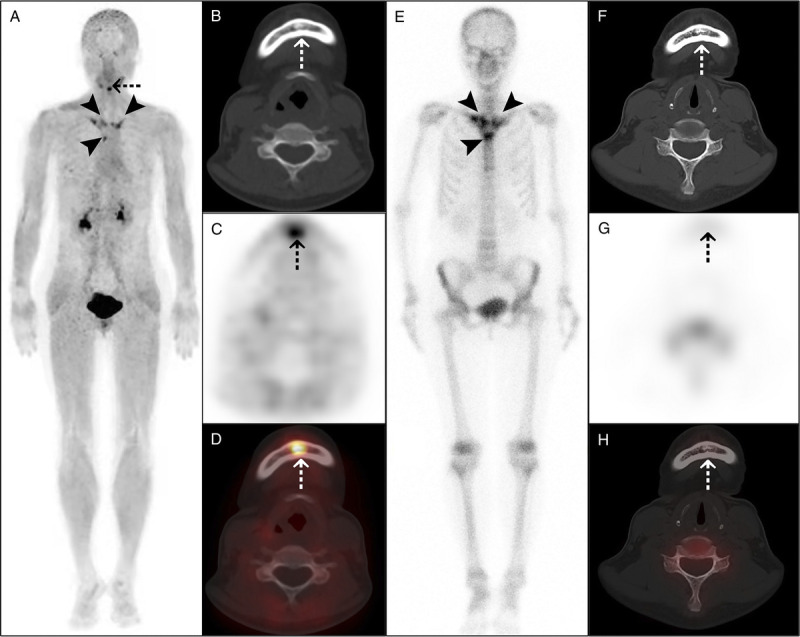
FIGURE 3.

68Ga-FAPI-04 PET/CT detected more osteoarticular lesions than 99mTc-MDP bone scan in a 53-year-old woman with SAPHO syndrome. The woman presented with anterior chest wall pain for 3 years. Laboratory examination showed elevated ESR (32 mm/h) and a positive ANA test. The maximum intensity projection (A) of 68Ga-FAPI-04 PET/CT showed increased tracer uptake in the bilateral sternoclavicular joints, bilateral anterior first ribs, and sternal angle (arrowheads). Moreover, PET/CT (A–D) showed osteosclerosis in the mandible, with increased tracer uptake (dotted arrows). 99mTc-MDP bone scan (E) showed increased tracer uptake in the above sites of anterior chest wall (arrowheads), resembling the bull's-head sign. However, 99mTc-MDP bone scan showed no abnormal bone metabolism in the mandible (F–H, dotted arrows).
