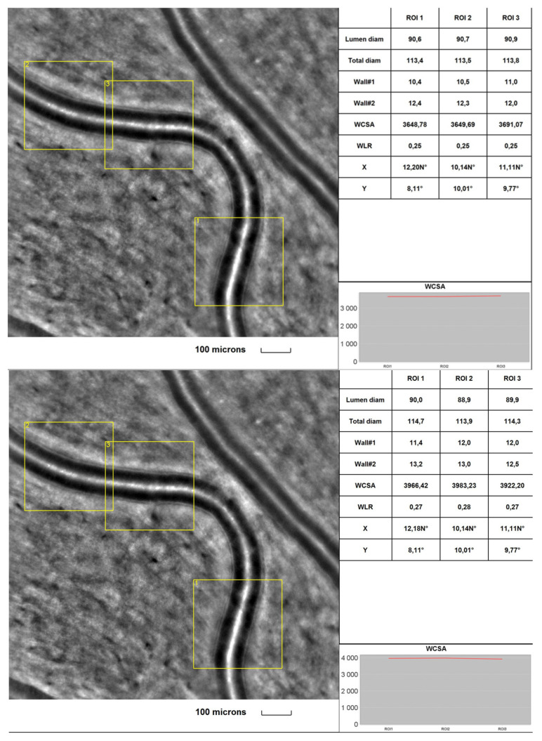
Figure 2.
Image of the retinal artery of a DR patient captured by rtx1TM AO retinal camera in three different acquisitions: ROI (region of interest), initially (upper) and after a 2-year observation time (lower). The charts present the following parameters: Lumen diam—lumen diameter; Total diam—total diameter; wall1 and wall2, WCSA—cross-sectional wall area; —WLR—wall-to-lumen ratio.

