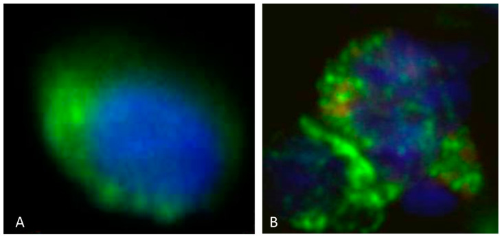
Figure 2.
(A): CTC cell isolated from the peripheral blood from a patient with diffuse intrinsic pontine gliomas (DIPGs). The CTC displays rounded morphology and is CSV positive (displayed by green fluorescence) and H3K27M negative. The nucleus is seen in blue. (B): CTC cell isolated from the peripheral blood in a patient with DIPG. The CTC displays rounded morphology and is CSV positive (displayed by green fluorescence) and H3K27M positive (displayed by red fluorescence). The nucleus is seen in blue. Scale indicates 10 µm.

