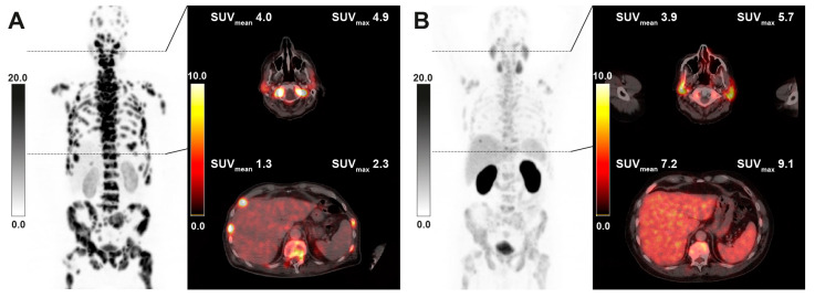
Figure 3.
Sample [68Ga]Ga-PSMA-11 PET/CT images of a 65-year-old patient with diffuse bone marrow involvement receiving 6 cycles of RLT with cumulative 53.5 GBq of [177Lu]Lu-PSMA-617. Maximum intensity projection (MIP) images (left) with axial fusion images showing the parotid gland and liver uptake at baseline (A) and after RLT (B). The decrease in tumor burden is associated with markedly increasing liver uptake, while salivary uptake remains nearly unchanged.

