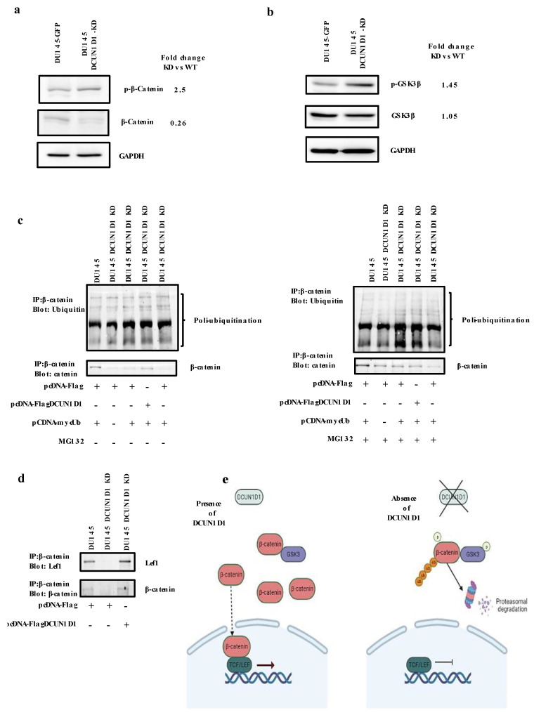
Figure 6.
Inhibition of DCUN1D1 deactivated the WNT signalling pathway. Protein extracts containing DU145-GFP and DU145 DCUN1D1-KD cell lines were analysed using Western blot analysis. Inhibition of DCUN1D1 (a) increased phosphorylation of β-catenin and reduced expression of total β-catenin and (b) increased the phosphorylated levels of Gsk-3β. The GAPDH was used as loading control. (c) Blockage of DCUN1D1 inhibits ubiquitination of β-catenin. DU145 DCUN1D1-KD and DU145-GFP cells were transfected with pCDNA Myc–Ub and pCDNA Flag–DCUN1D1 expression vector or parental vector and treated with proteasome inhibitor (MG132) for 4 h. Proteins were immunoprecipitated using anti-β-catenin antibody. Ubiquitinated β-catenin was detected by using anti-ubiquitin antibody. Re-expression of DCUN1D1 protein restores β-catenin expression. (d) Blockage of DCUN1D1 expression inhibited β-catenin/ Lef1 interaction. Proteins from DU145 and DU145 DCUN1D1-KD transfected with pCDNA Flag–DCUN1D1 cells were immunoprecipitated using anti-β-catenin antibody. The interaction between the proteins was detected by anti- Lef1 antibodies (e) Schematic representation of the DCUN1D1 pathway in PCa. Experiments were independently repeated three times and a representative image of an independent experiment is represented.

