Figure 7.
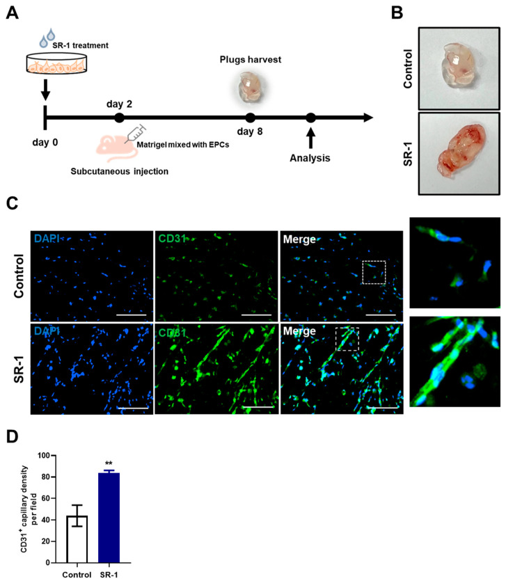
Effect of SR-1 on EPC-mediated angiogenesis. (A) Schematic diagram of an in vivo Matrigel plug assay. Matrigel-containing SR-1-treated EPC were subcutaneously injected into nude mice. (B) After 6 days, the plugs were collected and photographed. (C) For quantitative analysis of angiogenesis, endothelial cells were stained with CD31 (Green). The nucleus was stained by DAPI (Blue) (scale bar, 100 μm). The boxed regions are shown at a high magnification (×4) in the inset. (D) percentage of CD31+ capillary density was quantified. The data are presented as mean ± standard deviation. (** p < 0.01 vs. control, SR-1 1 μM treatment).

