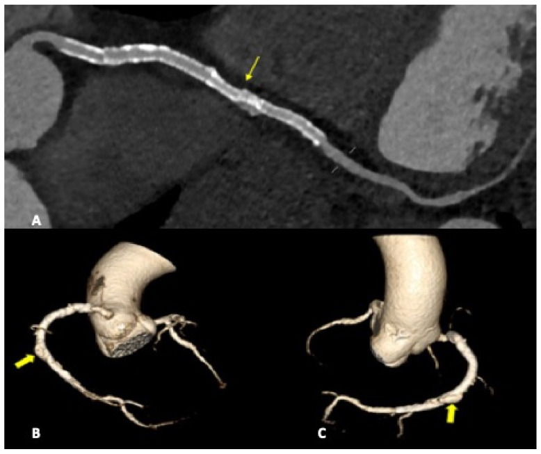
Figure 3.
CCTA, a curved planar reconstruction of RCA (A) and volume-rendering reconstruction of coronary tree (B,C). A: multiple stents in RCA can be seen. The most distal stent is misaligned (light arrow), with vascular dilatation and endoleak in the same vascular tract. (B,C): medium-contrast extravasation outside the stent in the distal RCA determines a constrast sacciform collection outside the stent meshes as an endoleak type III formation (heavy arrows).

