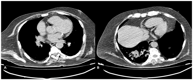
Figure 7.
Non-contrast-gated CT, axial planes: air presence in the RCA and its branches can be noted. Scan was performed in the immediate follow-up after a CT-guided lung-biopsy procedure, in a symptomatic (chest pain and dyspnea) patient. (A) air bubbles in proximal RCA can be seen. (B) Air is present in the distal RCA and posterior descendent artery.

