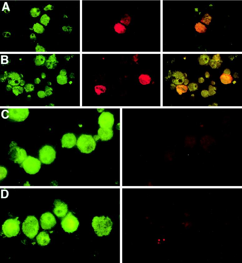
FIG. 2.
HHV-6 infects DC. DC were propagated from either adherent PBMC (A and C) or elutriated monocytes (B) in the presence of GM-CSF and IL-4, purified, and exposed overnight to HHV6Z29 at an MOI of 0.1. Excess virus was washed out, and cells were placed back into culture. Uninfected DC were cultured in parallel (D). The cells were cytospun onto glass slides 7 days after infection, fixed, and incubated with anti-CD1a MAbs (green [A to D]), anti-HHV-6 MAbs (red [A, B, and D]), or isotype control MAbs (red [C]). Yellow cells (A and B) represent CD1a+ DC productively infected with HHV-6.

