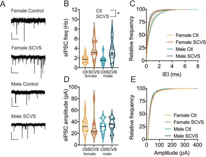
Figure 2: sIPSC frequency onto dopaminergic neurons is increased following SCVS in both male and female mice.
A. Representative traces of spontaneous IPSCs from dopaminergic neurons. B. Average sIPSC frequency from VTA dopaminergic neurons from control and SCVS mice. *p<0.05, 2-way ANOVA, main effect of stress. C. Cumulative distribution of sIPSC inter-event intervals. D. Average sIPSC amplitudes from VTA dopaminergic neurons from control and SCVS mice. E. Cumulative distribution of sIPSC amplitudes. n=10–11/group. Scale bars=50 pA, 1 s.

