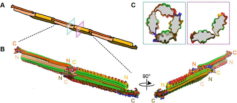
Figure 9. Filamentous multimer model for bacterial INPs.

A) A possible assembly of INP solenoids to form long fibres composed of antiparallel INP dimers (indicated by orange and yellow pairs). B) Dimers are formed along the tyrosine ladder, a previoiusly proposed dimerization interface. They are joined end to end by forming electrostatic interactions between negatively (red) and positively (blue) charged surfaces. All threonines are coloured light green, displaying the arrays of TxT WO-motifs. The termini of the INP solenoids are labeled N and C and coloured to match panel A. This illustration uses a manually flattened AlphaFold model of PbINP. C) Cross sections of the model at positions indicated in A. Monomers are rotated approximately 90° to each other and dimerized along their tyrosine ladders (purple). Toward their termini, a pair of dimers can be matched by oppositely charged electrostatic surfaces (teal).
