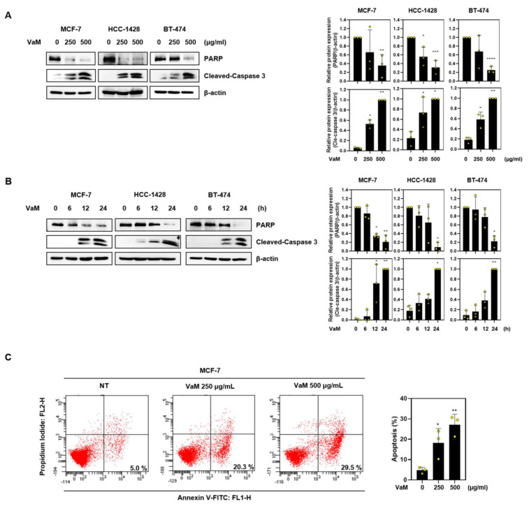
Figure 2.
Apoptotic effect of VaM on breast cancer cells. Cells were treated with VaM (A) at various concentrations for 24 h and (B) 500 μg/mL at the indicated time points. The protein expression of Pro-PARP and cleaved-caspase 3, which are apoptosis markers, was confirmed by immunoblotting. The data were quantified and compared to those of the untreated groups. (C) Flow cytometry was used to analyze Annexin V/PI-stained cells. * p < 0.05, ** p < 0.01, *** p < 0.001, and **** p < 0.0001 compared to the condition with zero treatment concentration. The results are presented in the bar chart as the mean ± SD of the three independent experiments. Yellow circles represent the results of three independent experiments.

