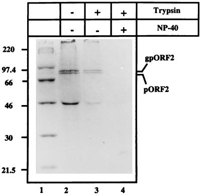
FIG. 4.
Microsomal localization of pORF2. COS-1 cells transfected with pSG-ORF2 were labeled with [35S]promix for 4 h and used to prepare the microsomal fraction as described in Materials and Methods. The microsomal fraction was then incubated in the absence (−) or presence (+) of 25 μg of trypsin/ml and/or 0.5% NP-40, on ice for 60 min. Following immunoprecipitation, the polypeptides were analyzed on an SDS–10% polyacrylamide gel, followed by fluorography. The positions of gpORF2 and pORF2 are indicated. Molecular size markers are shown (lane 1).

