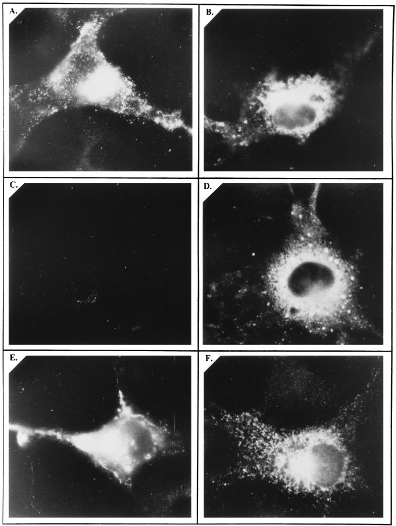
FIG. 7.
Cell surface and intracellular localization of pORF2 and its mutants. COS-1 cells transfected with pSG-ORF2 (A and B), pSG-ORF2[Δ2-34] (C and D), or pSG-ORF2[137,310,562] (E and F) were fixed with 4% paraformaldehyde–PBS and stained with rabbit anti-pORF2, followed by the goat anti-rabbit IgG-FITC conjugate. Antibody incubations were carried out in the absence (A, C, and E) or presence (B, D, and F) of 0.1% saponin for surface and intracellular staining, respectively. The stained cells were mounted in 20% glycerol and viewed and photographed with a fluorescence microscope. Representative views are presented.

