Figure 1.
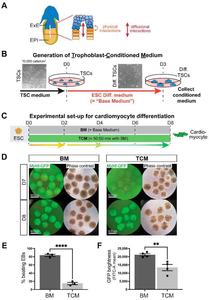
Trophoblast-conditioned medium affects cardiomyocyte differentiation from ESCs. (A) Diagram of an E6.5 mouse conceptus with a magnified view of the embryonic-trophoblast interface enabling possibilities for crosstalk. EPI = epiblast, ExE = extra-embryonic ectoderm. (B) Schematic depiction of the generation of trophoblast-conditioned medium (TCM). Trophoblast stem cells (TSCs) were exposed to ESC cell differentiation medium, also referred to as “base medium” (BM) that does not contain any growth factor supplements, for three days. Because of the absence of FGF, TSCs start to differentiate during this time. (C) Experimental set-up for cardiomyocyte differentiation time course from embryonic stem cells (ESCs) in either base medium (BM) or media consisting of 50% TCM: 50% BM, for brevity referred to as “TCM” in the subsequent figures. (D) Cardiomyocyte differentiation in embryoid bodies visualized by a cardiac-specific α-myosin heavy chain (MHC, Myh6) promoter-driven enhanced green fluorescent protein (GFP) construct shows the appearance of GFP-positivity at day 7 (D7) in BM but only at D8 in TCM. For visualization purposes, EBs were cultured in suspension culture from D2 onward. Data are representative of 12 independent biological replicates. (E) Percentage of beating embryoid bodies (EBs) at D8 of differentiation in EBs grown in suspension culture from D2 onward in BM or TCM. **** p < 0.0001, two-tailed Student’s t-test. (F) GFP intensity measured by flow cytometry as FITC-A mean in EBs at D10 of differentiation in BM or TCM. Data are displayed as the mean ± SEM. Statistical analysis was conducted by unpaired two-tailed Student’s t-test. ** p < 0.01.

