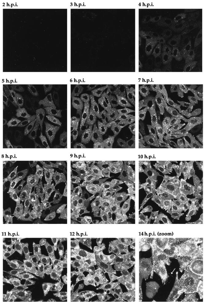
FIG. 6.
Live-cell analysis of GFP-22 localization during a high-multiplicity infection of 166v. Vero cells were infected with 166v at 10 PFU per cell and were examined every hour up to 14 h postinfection (14 h.p.i.) for GFP-22 fluorescence. The same settings for the confocal microscope were used at each time point. Extracellular fluorescent particles can be seen in the image taken at 14 h postinfection (arrow).

