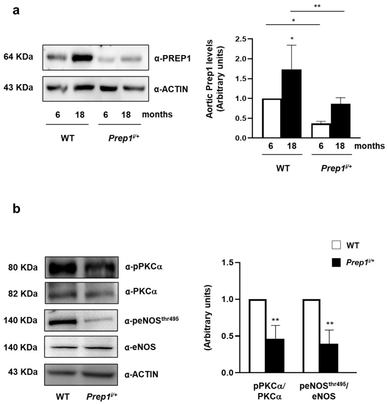
Figure 6.
The role of PREP1 on eNOS signaling in WT and Prep1i/+ mice. (a,b) Aortas from 6- to 18-month-old WT and Prep1i/+ mice were solubilized, and protein samples were analyzed by a Western Blot with PREP1, pPKCα, PKCα, peNOSThr495, eNOS antibodies. ACTIN antibody was used for normalization. Blots were revealed by ECL, and the autoradiograph was representative of three independent experiments and subjected to densitometric analysis. eNOS bands were taken from a parallel gel loaded with the same lysates. Asterisks denote statistical differences (* p < 0.05; ** p < 0.01). Arbitrary units are related to fold changes relative to WT.

