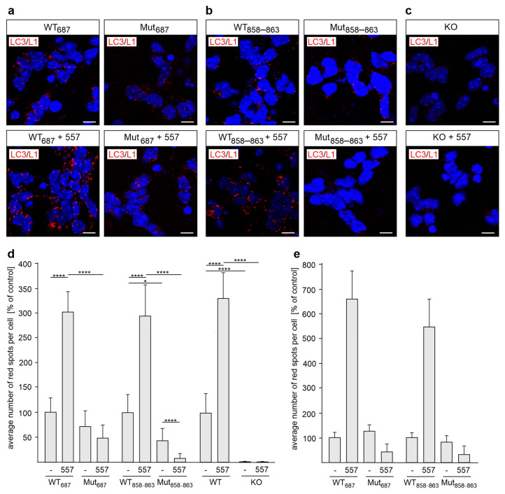
Figure 4.
LC3 interacts with L1-70 in cultured cerebellar and cortical neurons. Cultured cerebellar (a–d) and cortical (e) neurons from L1-70-lacking L1687 (Mut687) or L1858–863 (Mut858–863) male mutant mice (a–e) or L1-deficient (KO) male mice (d) and their male wild-type (WT) littermates (a–e) were treated without (-) or with L1 antibody 557 (557). The neurons were fixed and subjected to proximity ligation with mouse L1 antibody C-2 and a rabbit LC3 antibody. Nuclei are stained with DAPI (blue). (a–c) In the representative images, red spots indicate a close interaction of L1 with LC3. Scale bars: 10 μm. (d,e) Mean values + SD from three independent experiments (d) or one experiment (e) are shown for the average numbers of L1/LC3-positive spots per cell in cerebellar (d) or cortical (e) neurons (* p < 0.05, **** p < 0.001 one-way ANOVA with Dunn’s multiple comparison test).

