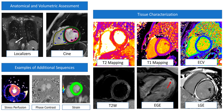
Figure 1.
Examples of CMR sequences in cardio-oncology. Anatomical localizers and cine sequences represent the core of CMR evaluation, allowing the detection of extra-cardiac abnormalities (white arrows) and volumetric and function assessment (multi-colored regions of interest). This basic protocol is also very quick (~20–30 min) and useful when dealing with uncompliant or sick patients or in serial surveillance scans. CMR can also detect myocardial oedema (T2 weighted images) and fibrosis (T1 mapping, ECV, EGE, and LGE). Information derived by tissue characterization has been demonstrated to be helpful in enhancing the sensitivity of cardiotoxicity diagnosis. In addition, EGE sequences are particularly helpful in detecting intracardiac thrombi (red arrow). According to clinical suspicion, the CMR protocol could be tailored to the patient and include additional sequences such as stress perfusion imaging (to assess large and small vessel coronary disease), phase contrast (for flows and valvular function evaluation), and strain sequences. CMR: cardiovascular magnetic resonance. ECV = extracellular volume. EGE = early gadolinium enhancement. LGE = late gadolinium enhancement.

