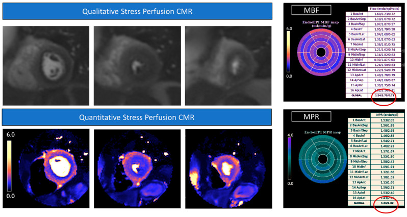
Figure 2.
Stress perfusion Cardiovascular Magnetic Resonance. Raw perfusion images showed a diffuse hypointense «darker» area in all the coronary territories representing a myocardial perfusion defect during drug-induced (adenosine) hyperemia. The diffuse subendocardial perfusion defect is also highlighted in the colored perfusion maps, where hypoperfused areas are shown in blue. The quantitative analysis showed significantly reduced myocardial blood flow (MBF) values, particularly in the subendocardium (1.24 mL/min/g), and concomitant reduced subendocardial myocardial perfusion reserve (MPR) values (1.36).

