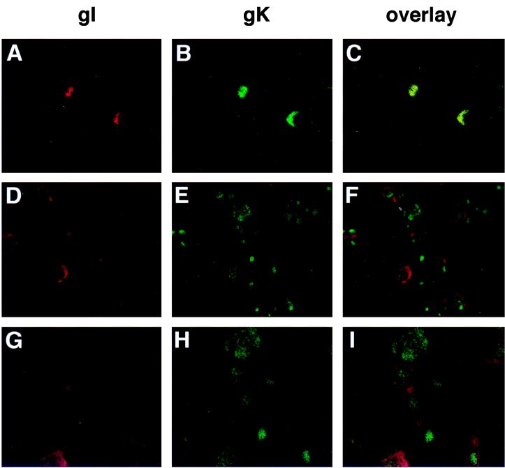
FIG. 4.
Immunofluorescence analysis of VZV gI and gK distribution. Melanoma cell monolayers growing on glass coverslips were inoculated with VZV-infected cells. The cells were fixed and either not permeabilized (A to C) or permeabilized (D to I). The cells were incubated with primary mouse anti-gI and rabbit anti-gK sera before incubation with Texas red-conjugated goat anti-mouse IgG and FITC-conjugated goat anti-rabbit IgG. Cells were examined with a Molecular Dynamics MultiProbe 2010 laser scanning confocal microscope. Magnification, ×200 (A to F) and ×400 (G to I).

