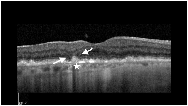
Figure 2.
Incomplete retinal pigment epithelium outer retinal atrophy (iRORA). Foveal cut of an SD-OCT (Heidelberg Spectralis) image of an 83-year-old woman with a visual acuity of 20/80. The arrows point to the presence of a hyporeflective wedge in the Henle fiber layer (HFL). The star is adjacent to an area of choroidal hypertransmission <250 µm.

