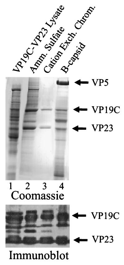
FIG. 3.
SDS-polyacrylamide gel electrophoresis of fractions obtained during triplex purification (lanes 1 to 3). For comparison, HSV-1 B-capsid proteins are shown in lane 4. The upper panel shows the gel after staining with Coomassie blue, while the bottom panel shows the results of Western immunoblotting of the VP19C-VP23 region in a comparable gel after staining with rabbit polyclonal antibodies specific for VP19C and VP23. Amm., ammonium; Exch. Chrom., exchange chromatography.

