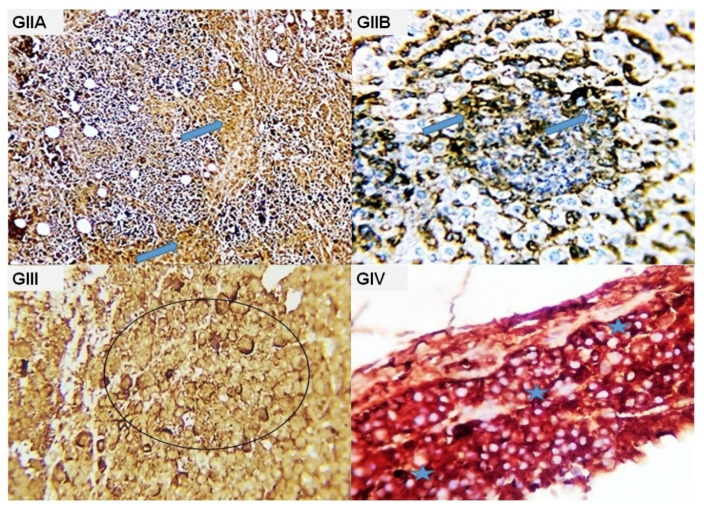
Figure 6.
Photomicrograph from the tumor mass of different experimental groups immune-stained with CK. (GIIA, GIIB): Mark brownish cytoplasmic stainability of the malignant cells (blue arrows), (GIII): massive necrosis of the tumor cells with negative staining reaction. Some cells showed hazay cytoplasmic membrane brownish staining reaction (black circle); (GIV): remnants of tumor mass were seen with positive cytokeratin cytoplasmic staining reaction in most of the remaining cells (blue stars).

