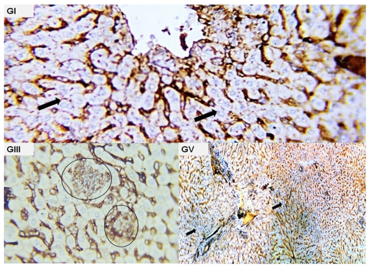
Figure 7.
Photomicrograph from the liver of different experimental groups immune-stained with CK. (GI): normal hepatocytes free from any malignant cells (black arrow); (GIII): focal aggregates (deposit) of weak positive malignant cells (black circles); (GV): liver parenchyma appeared negative for any of metastatic cells, no positive reactivity for the used marker was detected (black arrow).

