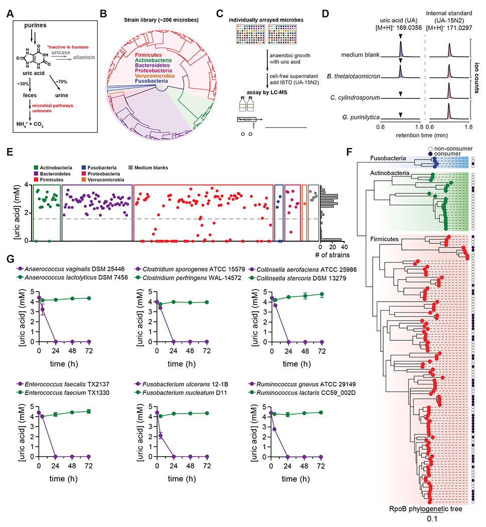
Figure 1. Anaerobic uric acid metabolism is widespread among human gut bacteria.

A) Overview of purine metabolism in humans. B) Phylogenetic distribution of human gut bacteria in the strain library used for this study. C) Overview of experimental approach to screen for uric acid metabolism. D) Extracted ion chromatograms for uric acid and the uric acid internal standard (ISTD; [15N2]-uric acid) in medium blank and after incubation with a non-consumer (B. thetaiotaomicron) and two known purine-consuming bacteria (C. cylindrosporum and G. purinilytica). E) Results from uric acid screen in rich medium, grouped by phylum. Each dot represents a single bacterial strain. The frequency of strains is shown on the right of the plot. F) Phylogenetic distribution of uric acid consuming bacteria within the Actinobacteria, Fusobacteria, and Firmicutes phyla. Dark purple dots represent strains that consume >50% of the uric acid. Only those strains for which assembled genomes are available were included. G) Uric acid consumption in closely related bacteria during growth in rich media. For D and E, data represent the results from a single experiment. For G, data represent the means ± standard deviations of n = 3 biological replicates.
