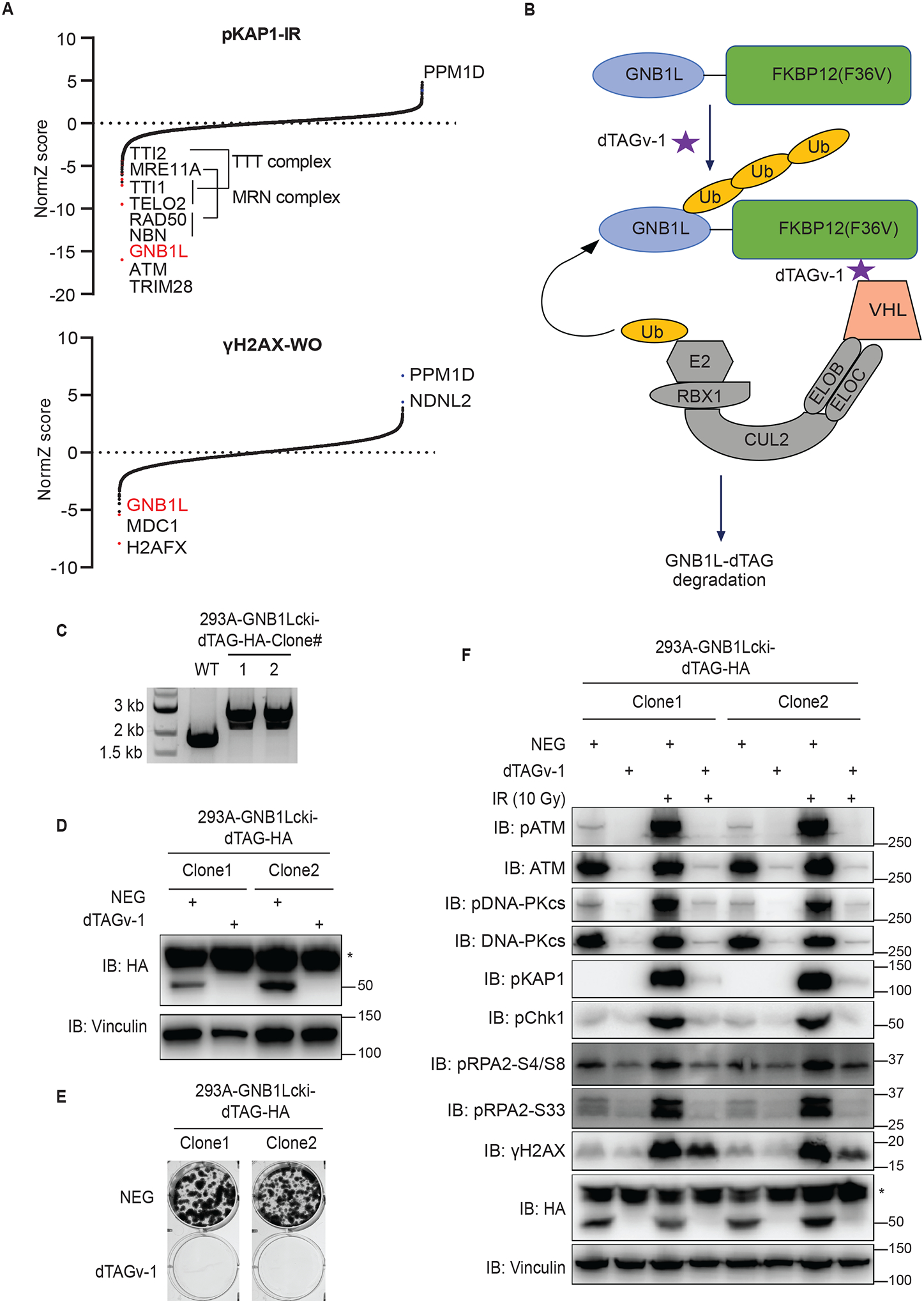
Figure 5. GNB1L depletion significantly reduces both basal and IR-induced DDR signaling.

(A) Results of DrugZ analysis in FACS-based screens performed with antibodies recognizing endogenous pKAP1 or γH2AX.
(B) Schematic of the dTAG system using VHL-recruiting dTAGv-1 to promote the degradation of GNB1L-dTAG protein.
(C) PCR validation of GNB1Lcki-dTAG clones of HEK293A cells.
(D) Western blot validation of the GNB1Lcki-dTAG cell lines.
(E) Clonogenic survival of GNB1L-dTAG cells in the presence of dTAGv-1 or dTAGv-1-NEG (NEG).
(F) Immunoblots of DDR signaling proteins in GNB1L-dTAG cells. Cells were treated with dTAGv-1 or dTAGv-1-NEG for 3 days and followed by exposure to IR (10 Gy, allowed recovery for 1 h) or mock treatment.
