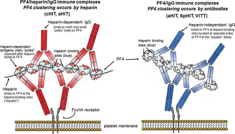
Fig. 2.

Heparin-dependent versus heparin-independent antibodies. Left : Platelet activation by heparin-dependent antibodies. Shown is a small immune complex composed of 7 PF4 molecules bound together (“clustered”) by a lengthy heparin molecule, and 4 IgG antibodies. PF4 is a highly charged (cationic) molecule; thus, PF4 clustering requires (polyanionic) heparin. Binding of the globular tetrameric protein, PF4, to heparin results in formation of antigen sites at the north and south “poles” (indicated in red). The IgG Fc “tails” bind to the platelet FcγIIa receptors; crosslinking of the FcγIIa receptors results in strong platelet activation. Right : Platelet activation by heparin-independent antibodies. Shown is a small immune complex composed of 7 PF4 molecules and 4 IgG antibodies. The heparin-independent antibodies bind to the heparin-binding site on PF4 (“equator”). In this case, PF4 clustering is caused by the IgG antibodies themselves. This is the general mechanism for VITT antibodies as well as those heparin-independent antibodies within aHIT sera. HIT, heparin-induced thrombocytopenia; VITT, vaccine-induced immune thrombotic thrombocytopenia.
