FIG. 1.
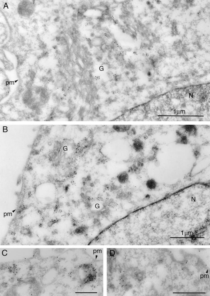
Immunoelectron microscopy of Us9-EGFP. Thawed ultrathin sections of PK15 cells stably expressing a Us9-EGFP fusion protein were stained with a polyvalent rabbit Us9 antiserum followed by protein A conjugated to colloidal gold (10 nm). Low-magnification views illustrate the predominant staining of Golgi-associated membranes and vesicular structures (A and B). No staining of the nuclear membrane is observed. Low levels of Us9-EGFP can be detected on the plasma membrane (C and D). Vesicular structures throughout the cytoplasm were seen singularly, in clusters, or in structures reminiscent of endosomes (C). In some instances, Us9-EGFP-containing vesicles are observed close to the plasma membrane and seem to be in the process of fusing with the plasma membrane (D). Magnification bars in panels C and D represent 500 nm. pm, plasma membrane; G, Golgi apparatus; N, nucleus.

