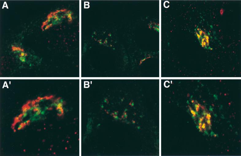
FIG. 3.
Colocalization of Us9 with Golgi and TGN markers. PK15 cells grown on glass coverslips were transfected with plasmids pBB14 (A and B) and pAB7 (C). After 36 h of transfection, the cells were fixed and stained for Us9 and mannosidase II (A), for Us9 and p115 (B), and for Us9 and TGN38 (C). Us9 was detected either by GFP fluorescence (A and B) or with a fluorescein isothiocyanate-conjugated secondary antibody (C) which fluoresces green. Mannosidase II (A), p115 (B), and TGN38 (C) were visualized with an Alexa 568-conjugated secondary antibody which fluoresces red. High-magnification views are shown in panels A′, B′, and C′.

