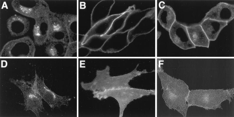
FIG. 7.
Transfection of Us9 constructs. PK15 cells grown on glass coverslips were transfected by the calcium phosphate method with pBB14 (A and D), pAB35 (B and E), or pAB37 (C and F). At 72 h posttransfection, the intracellular localization of the various Us9-EGFP fusion proteins was detected by confocal microscopy (A to C). The cell surface of the transfected cells was visualized by fluorescence microscopy (D to F).

