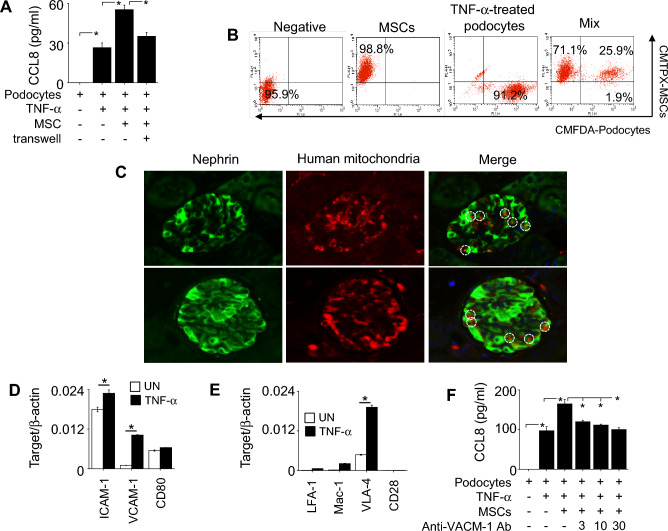
Figure 3.
Effect of MSCs on CCL8 production by podocytes. (A) Podocytes (1 × 10⁵ cells/well) were loaded into the lower wells of transwell plates. MSCs (0.1 × 10⁵ cells/well) were added to the upper wells to prevent contact or to the lower wells to allow contact with podocytes. After incubation for 24 h, CCL8 level in the medium was determined by ELISA. (B) Binding rates of CMTPX-labeled MSCs (1 × 10⁶ cells/tube) and CMFDA-labeled podocytes (1 × 10⁶ cells/tube) were analyzed by flow cytometry. Representative dot plots are shown. (C) Mice were intravenously injected with human MSCs (1 × 10⁶ cells/mouse) at the age of 12 weeks (n = 6). Kidneys were isolated 24 h later. Kidney sections were stained with rabbit anti-mouse nephrin antibody conjugated with GFP (green) and mouse anti-human mitochondria antibody conjugated with Alexa Fluor 647 (red). Representative contacts are shown in circles. (D) The levels of ICAM-1, VCAM-1, and CD80 mRNAs in TNF-α-treated podocytes were determined by RT-qPCR. (E) The levels of LFA-1, Mac-1, VLA-4, and CD28 mRNAs in TNF-α-treated MSCs were determined by RT-qPCR. (F) Podocytes were pre-treated with anti-VCAM-1 antibody at 3–30 ng/ml for 2 h. After washing, podocytes (1 × 10⁵ cells/well) were co-cultured with MSCs (0.1 × 10⁵ cells/well). After incubation for 24 h, CCL8 level in the medium was determined by ELISA. TNF-α was used at 50 ng/ml in all experiments. RQ, Relative quantitation. UN, Untreated. n = 3 in panels of (A–B) and (D–F). *p < 0.01.

