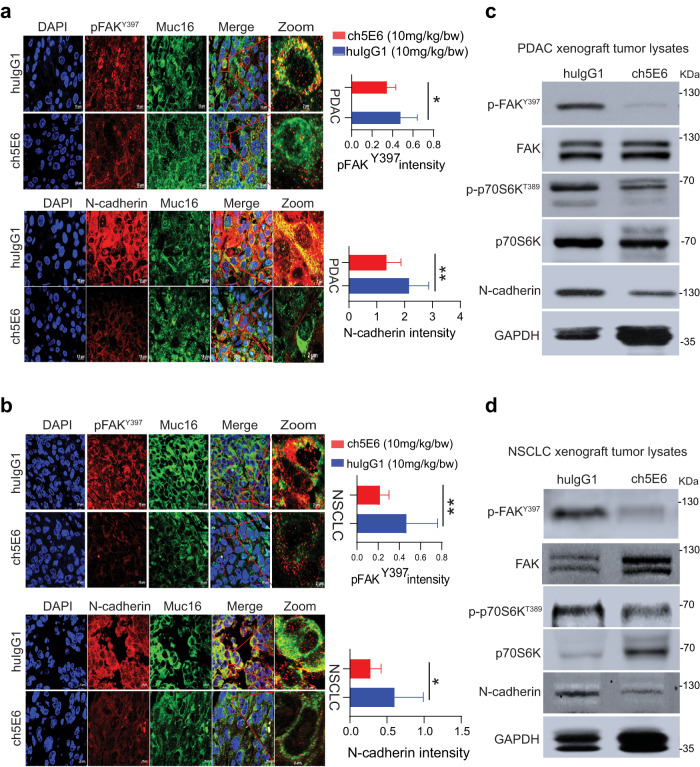
Fig. 6. Inhibition of EMT by ch5E6 is validated in PC and NSCLC cell line-derived xenografts.
a Immunofluorescence analysis showing a decrease in pFAK(Y397) and N-cadherin expression in xenograft tumors of SW1990 cells treated with ch5E6 compared to isotype control mAb huIgG1 group (n = 6–8 fields/tissue: three animals). The data was plotted for changes in fluorescence intensity using GraphPad Prism 9 and is shown in parallel. Nuclei were stained with DAPI. b Immunofluorescence analysis of ch5E6 treated SW1573 cell line-derived xenografts showing a reduction in pFAK(Y397) and N-cadherin levels compared to isotype control mAb huIgG1 group (n = 6–8 fields/tissue: 3 animals). Scale bar, 10 µm; magnified images, 2 µm. No significant changes in the intensity of MUC16 were seen in the ch5E6 treated versus isotype control tumors derived from both cancers. c, d Immunoblot analysis of ch5E6 treated PDAC and NSCLC tumor lysates showing a substantial decrease in phosphorylated levels of FAK(Y397), p70S6K(T389) and N-cadherin as compared to huIgG1 treatment. Error bars indicate SEM. Scale bar, 400 μm; magnified images, 100 μm; *P < 0.05; **P < 0.01.

