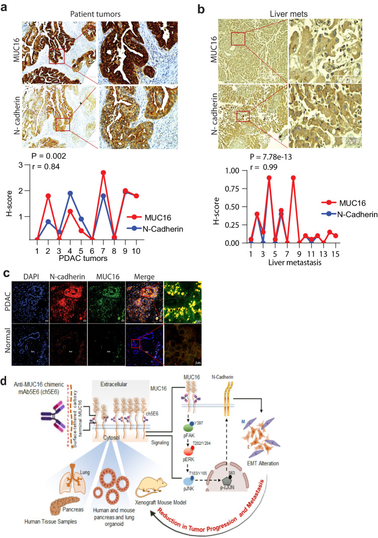
Fig. 7. MUC16 and N-cadherin are clinically correlated in patient tumors.
a, b Representative images and quantitative illustration of immunohistochemical analyses demonstrate a strong positive correlation between MUC16 and N-cadherin (R = 0.84) in both primary PC tumors (n = 10) and liver metastasis (R = 0.99) samples (n = 8). Scale bar, 400 µm; magnified images, 100 µm. *P < 0.05; **P < 0.01. c Representative images of immunofluorescence analysis showing coexpression of MUC16 (green) and N-cadherin (red) in primary PDAC tumors compared to no MUC16 and N-cadherin in normal pancreatic sections. Scale bar, 20 µm; magnified images, 5 µm. d Schematic diagram representing ch5E6 induced downregulation of MUC16 mediated EMT resulting in its anti-tumor potential in PC and NSCLC. Overall, anti-MUC16 chimeric mAb5E6 (ch5E6) binds to the cell surface-tethered domain of MUC16, interferes with oncogenic pFAK/p70S6K/N-cadherin signaling associated with MUC16-mediated EMT, and reduces tumor burden in both PC and NSCLC models.

