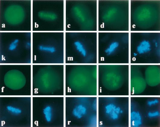
FIG. 6.
Mapping EBNA-1 chromosome binding sites in living cells. HeLa cells were transfected with plasmids encoding EGFP or various EBNA-1 derivatives fused to the EGFP protein: EGFP (a), EBV EBNA-1 (b), HVP EBNA-1 (c), M13 (d), M21 (e), M9 (f), M10 (g), M11 (h), M6 (i), and M7 (j). Cells were grown on cover slides, stained with Hoechst 33342, and observed in phenol red-free medium by low-light fluorescence microscopy either at 430 to 480 nm (EGFP) (a to j) or at 365 nm (Hoechst) (k to t).

