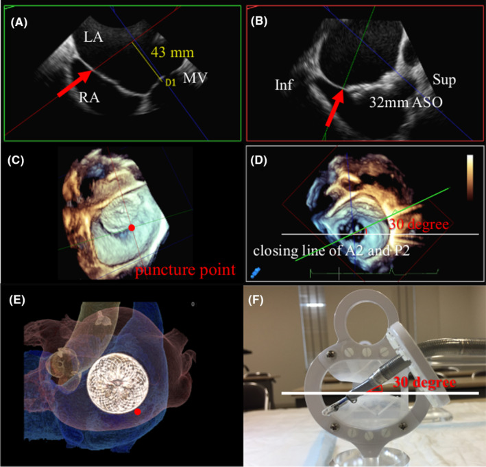
FIGURE 2.

(A) Multiplanar reconstruction showing the best puncture point (red arrow) within the fossa ovalis on a four‐chamber view. (B) Bicaval view showing the best puncture point (red arrow) out of the ASO device. (C) Estimated best puncture point (red dot) on 3D volume rendering view. (D) Estimated puncture point showing a 30° anterior side from the A2 and P2 closing line. (E) CT enface view showing the puncture point (red dot) correlated with TEE volume rendering view. (F) Demo device showing a nearly 30° approach angle. ASO, Amplatzer Septal Occluder; CT, computed tomography; Inf, inferior; LA, left atrium; MV, mitral valve; RA, right atrium; Sup, superior; TEE, transoesophageal echocardiography.
