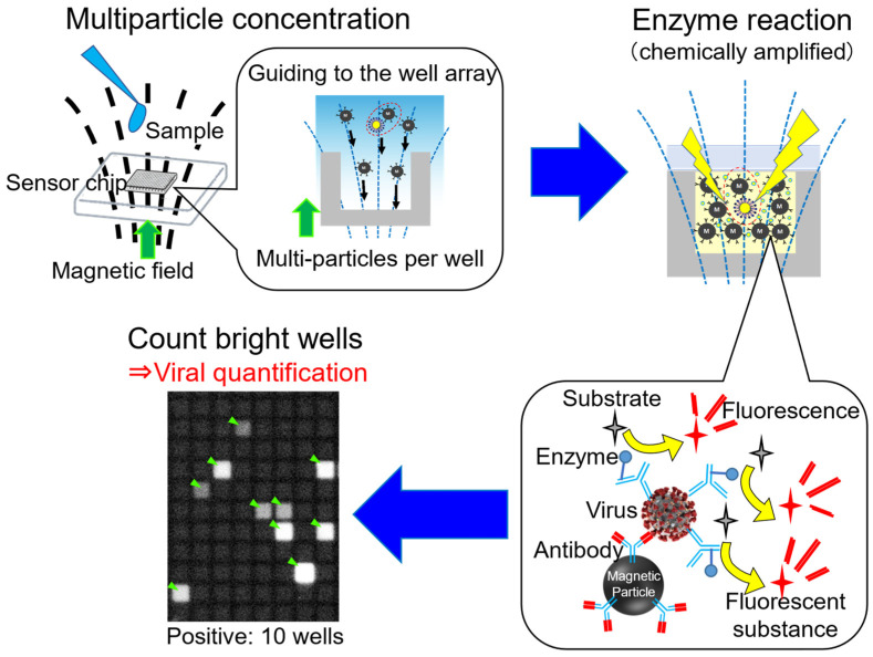
Figure 8.
Schematic diagram of MCIDA. After capturing the targets by antibody-modified magnetic beads, bead–target–enzyme immunocomplexes are applied to microwells. Different from the digital ELISA, many immunocomplexes and beads are introduced in each microwell. After sealing and enzyme reaction, the sealed microwell array is observed, and we counted how many wells indicate fluorescence. Green arrows indicate the direction of the magnetic field. Yellow arrows indicate the reaction of the substrates changing to the fluorescent substance.

