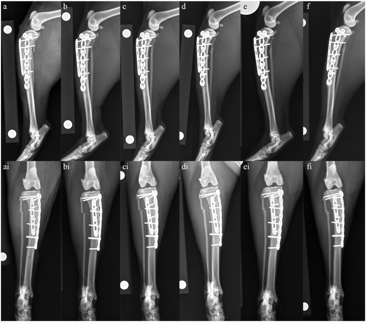
Figure 3.
Mediolateral and craniocaudal radiographs of the left tibia taken (a and ai) immediately postoperatively, (b and bi) 4 weeks postoperatively, (c and ci) 8 weeks postoperatively, (d and di) 14 weeks postoperatively, (e and ei) 22 weeks postoperatively and (f and fi) 17 months postoperatively. (a,ai) Immediately postoperatively, anatomic alignment and near-anatomic reduction have been achieved. Implant placement is appropriate with a 1.5 mm six-hole LCP placed cranially and a 1.5/2.0 mm split-T seven-hole LCP placed medially. (b,bi) At 4 weeks postoperatively, there is minimal evidence of healing at the tibial or fibula fracture sites. Anatomic alignment and near-anatomic reduction have been maintained and there is no evidence of implant-associated complications. (c,ci) At 8 weeks postoperatively, the tibial fracture line is slightly narrower cranially than at the 4-week re-check, and there is evidence of early callus formation caudally and medially. There is also evidence of early remodeling of the fibula fracture. Alignment and apposition have been maintained and implants remain static. (d,di) At 14 weeks postoperatively, there is progressive narrowing of both the tibial and fibula fracture lines when compared with the radiographs taken at 8 weeks postoperatively and evidence of moderate callus formation. Alignment and apposition have been maintained and implants remain static. (e,ei) At 22 weeks postoperatively, clinical union of the tibial fracture has been achieved with no signs of implant-associated complications. There is an oligotrophic nonunion of the fibula. (f,fi) At 17 months postoperatively, radiographs remain static when compared with those taken at 22 weeks postoperatively

