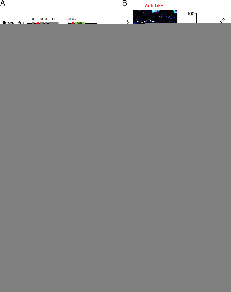
Figure 2.
c-Fos protects knee cartilage in experimental OA. (A) Targeting strategy and structure of the floxed/deleted c-Fos allele. Cre expression results in deletion of exons 2–4 and expression of nuclear EGFP under the control of the c-fos promoter. Coding areas (exon 1–4) are depicted in grey boxes. Experimental procedure and timeline to delete c-fos in chondrocytes and experimentally induced cartilage damage (DMM). Tamoxifen was injected intraperitoneally at two time points (2.5 and 9 weeks of age, 2 mg/mouse/day, 5 consecutive days) and mice were subjected to DMM/sham at 10 weeks of age and knee joints analysed 8 weeks post surgery. (B) Analysis of c-Fos deletion by anti-GFP immunofluorescence (red) in c-FosWT and c-FosΔCh mice articular cartilage at 10 weeks of age. The left panels are representative images of GFP-positive articular chondrocytes before surgery. (nuclei counterstained with DAPI) while % GFP-positive articular chondrocytes are plotted on the right. Articular cartilage is depicted with white dashed lines. (C) Representative images of safranin O/fast green staining of knee joints from c-FosWT mice and c-FosΔCh mice 8 weeks post surgery. (D) Quantification of cartilage damage on the medial side. (E) Relative cartilage area quantified by ImageJ analysis. Bar graphs and plots represent or include mean±SD, respectively. *p<0.05, **p<0.01, and ***p<0.001. Statistical differences between groups were analysed by non-parametric Mann-Whitney test in B and by two-way ANOVA with Bonferroni post hoc analysis in D and E. ANOVA, analysis of variance; DAPI, 4′,6-diamidino-2-phenylindole; DMM, destabilisation of the medial meniscus; EGFP, enhanced green fluorescent protein; OA, osteoarthritis; OARSI, Osteoarthritis Research Society International; TAM, Tamoxifen.

