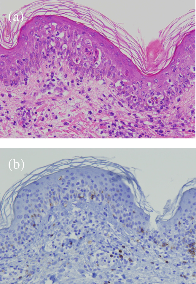
FIGURE 2.

(a) Hematoxylin and eosin staining showed keratinocyte necrosis and inflammatory cell infiltration, with mild lymphocytes infiltration from the epidermal layer to the shallow dermis. (b) Immunohistochemical staining showed CD8‐positive T cells infiltration from the epidermal layer to the shallow dermis.
