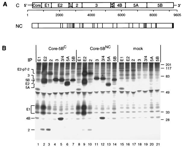
FIG. 1.
(A) Structure of the HCV genome and comparison of the C and NC polyproteins. (A) Schematic representation of the HCV genome organization, with the structural proteins core to E2, p7, and the nonstructural proteins NS2 to NS5B shown at the top. Numbers below refer to the nucleotide positions of our HCV isolate. Amino acid deviations of the NC polyprotein from the C sequence are indicated by vertical lines. (B) Expression of the complete ORFs of the NC and C isolates in BHK-21 cells and detection of cleavage products. Cells infected with the vTF7-3 vaccinia virus recombinant were transfected with plasmids directing expression of the complete polyprotein of the NC or C genome. After metabolic radiolabeling with [35S]methionine-cysteine, HCV-specific proteins were isolated from the cell lysate under nondenaturing conditions by immunoprecipitation (IP) using antisera with specificities given above the lanes. Identification of HCV-specific proteins and the positions of protein molecular mass standards (in kilodaltons) are given on the left and right, respectively.

