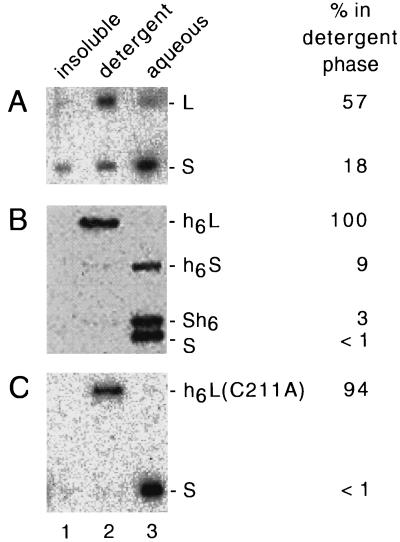
FIG. 4.
Triton X-114 phase separation of delta proteins. As described in Materials and Methods, samples containing different forms of the delta proteins were separated into three fractions (insoluble, detergent, and aqueous; lanes 1 to 3, respectively) and then assayed by gel electrophoresis and immunoblotting. (A) Nonidet P-40-disrupted particles from the serum of an HDV-infected woodchuck. (B and C) Phase separations for different combinations of purified delta proteins, as indicated on the right. The immunoblots were quantitated to determine for each delta protein the fraction of the total soluble protein detected in the detergent phase; the results are indicated at the far right.

