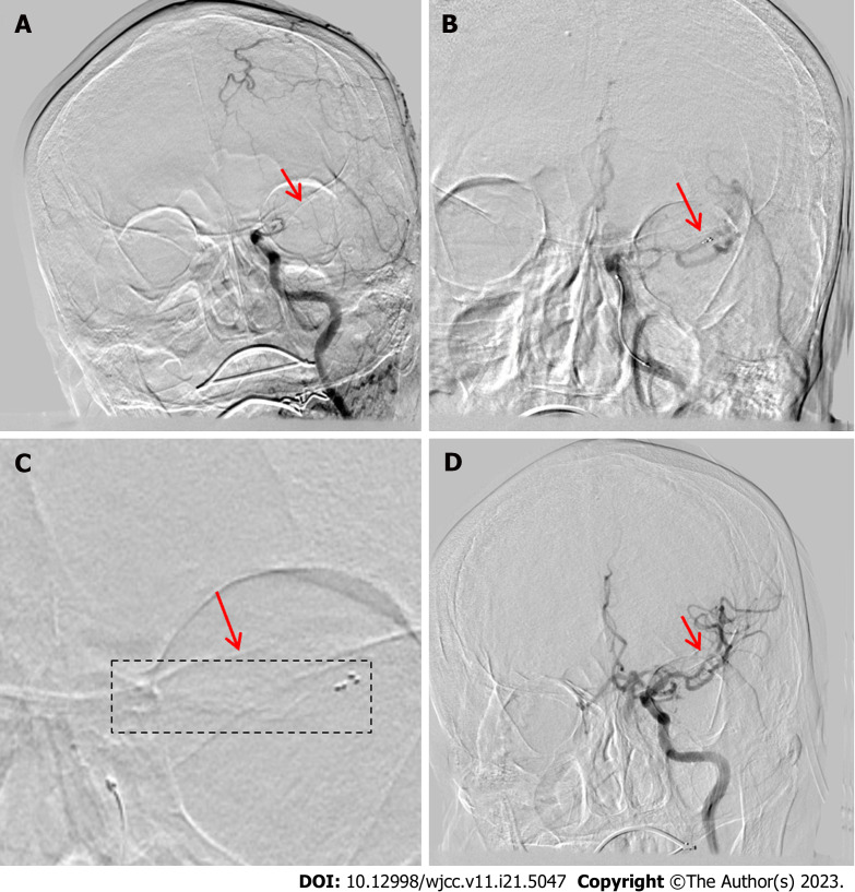
Figure 4.
Operative procedure: Patient 4. A: Digital subtraction angiography revealing left distal internal carotid artery (ICA) occlusion with poor blood flow compensation (arrow); B: Solitaire AB (6 mm × 30 mm) stent was deployed at the distal left middle cerebral artery-M1 segment (arrow); C: Stent imaging can be observed (arrow); D: Second (repeat) angiogram revealing good recanalization of the left ICA after the removal of the thrombi (arrow).

