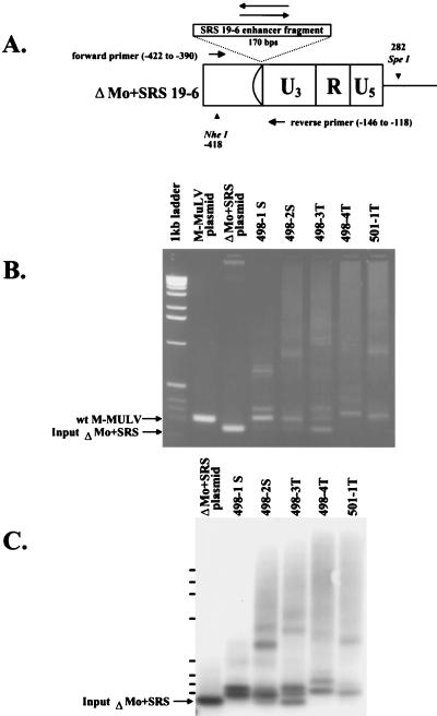
FIG. 4.
PCR analysis of proviral enhancers in ΔMo+SRS M-MuLV-induced tumors. (A) A diagram of the ΔMo+SRS+ or ΔMo+SRS− M-MuLV LTRs is shown, along with the locations of the oligonucleotide primers used to amplify the proviral LTRs from tumor DNAs. (B) PCR products from several different ΔMo+SRS M-MuLV-induced tumors were analyzed by agarose gel electrophoresis (2% agarose) and stained with ethidium bromide. Plasmids containing either wild-type (wt) M-MuLV or input ΔMo+SRS M-MuLV DNA provided size marker controls for the PCR amplification products, as shown to the left of the gel. Although some tumor DNAs yielded PCR fragments of the expected size, all tumors gave one or more enhancer-specific fragment of increased size. (C) Southern blot hybridization with an SRS enhancer-specific probe of a gel similar to the one in panel B is shown.

