
Figure 6.
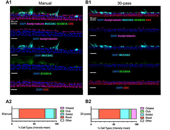
Cellular composition of manual seeding and bioprinted (30-pass) nasal epithelium. (A1) and (B1) and representative immunofluorescent images of differentiated hNECs characterized by major epithelial cell markers: basal (CK5, red), goblet (MUC5AC, cyan), club (SCGB1A1, green) and ciliated cells (acetylated α-tubulin, magenta) and nuclei (DAPI, blue). (A2) and (B2). Quantification of epithelial cell types in nasal ALI cultures by histocytometry (n = 1).