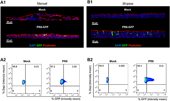
Figure 8.

Primary human nasal epithelial ALI cultures permissive to PR8-GFP influenza infection. Representative IF images of nasal ALI from (A1) manual seeding and (B1) bioprinting (30-pass) following 24 h exposure to uninfected control (Mock) or influenza virus (2.5 × 10⁵ pfu PR8-GFP); nuclei (DAPI, blue), phalloidin (actin filament, red), and GFP expressing influenza virus to reveal effective viral replication (GFP, green). Scale bar 50 μm, in white on the left corner. Representative histocytometry dot plots of (A2) manual seeding ALI and (B2) high-throughput bioprinted ALI with Mock and PR8-GFP conditions, showing the intensity mean for cell populations positive for DAPI (Y axis) and virus reporter GFP (X axis).
