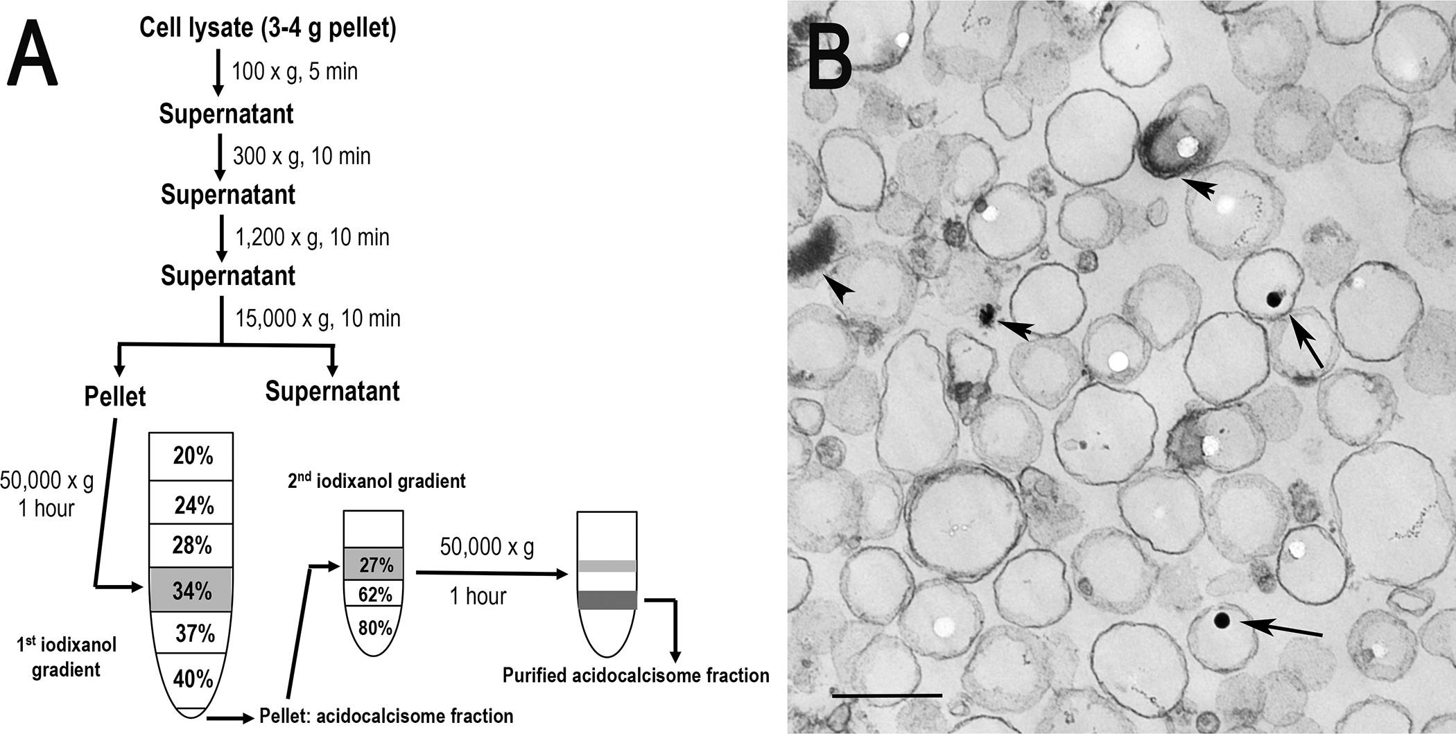
Fig. 1.

Subcellular fractionation of acidocalcisomes. (a) Trypanosome lysates are obtained by grinding with silicon carbide, decanting by low speed centrifugation to eliminate debris and silicon carbide, and centrifuging at 15,000 × g for 10 min to isolate the organellar fraction that is applied to the 34% step of a discontinuous iodixanol gradient. After centrifugation at 50,000 × g for 1 h, the pellet is resuspended and applied to the 27% step of a second iodixanol gradient and centrifuged at 50,000 × g for 1 h. Aliquots from each fraction are used for enzymatic assays and Western blot analyses. (b) Electron microscopy of acidocalcisome fraction prepared by the iodixanol procedure (fraction 5). Scale bar = 0.2 μm. Arrows and arrowheads show electron-dense material inside acidocalcisomes (reproduced from Huang et al. (2014) (ref. 3) with permission from the Public Library of Science).
