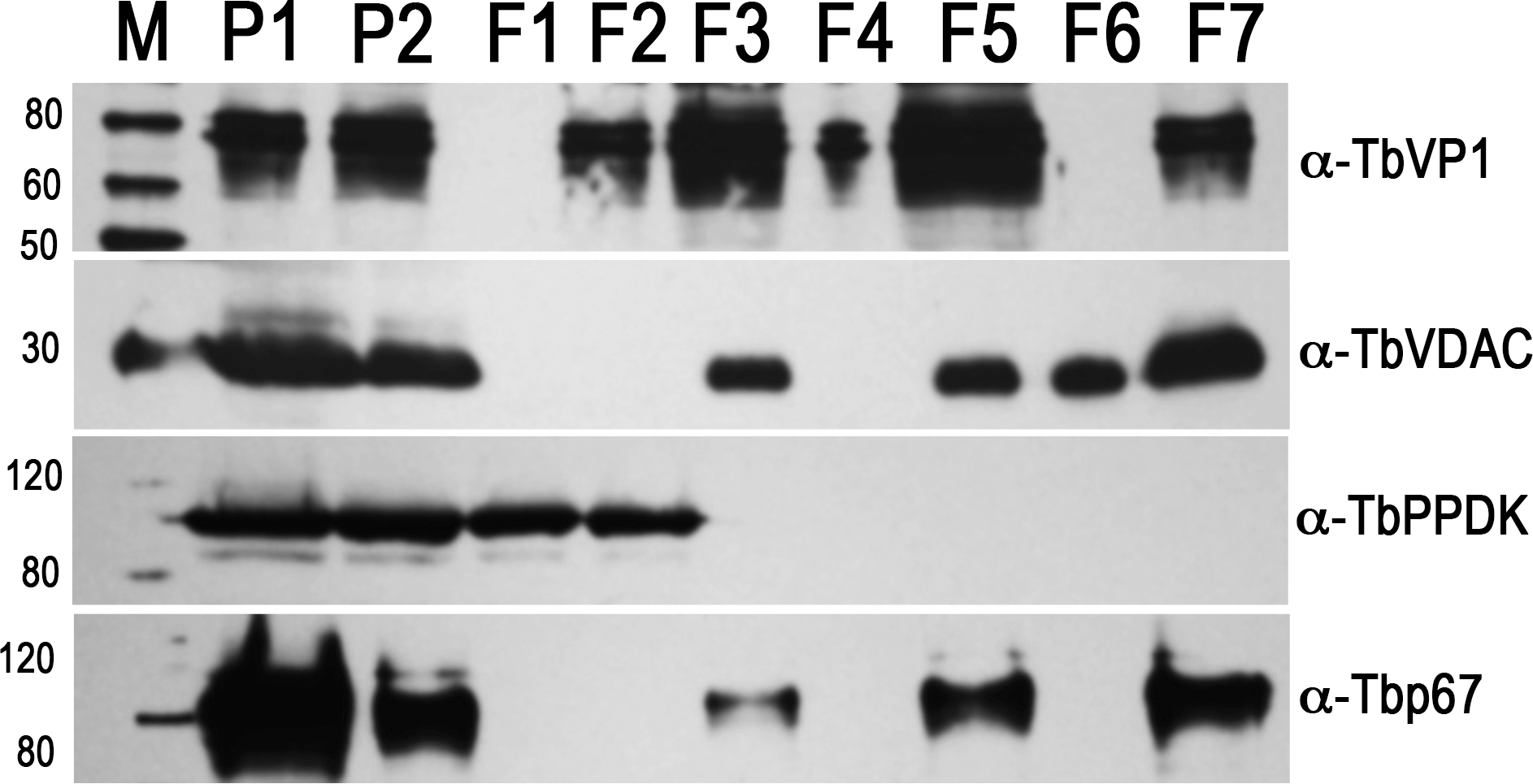
Fig. 3.

Western blot analyses of subcellular fractions using antibodies against acidocalcisome marker TbVP1, mitochondrial marker voltage-dependent anion channel (TbVDAC), glycosomal marker pyruvate, phosphate dikinase (TbPPDK), and lysosome marker Tbp67. P1, the 15,000 x g pellet (30 μg); P2, the first gradient pellet (2 μg); F1 to F7, the second gradient fractions (2 μg each). M, Magic Marker protein standards (reproduced from Huang et al. (2014) (ref. 3) with permission from the Public Library of Science).
