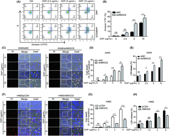
FIGURE 1.

AMIGO2 suppressed pyroptosis induced by cisplatin. (A) A549 cells transfected with AMIGO2 siRNA were treated with graded concentrations of cisplatin (DDP) for 48 h, and subjected to flow cytometric analysis after dual staining with Annexin V‐FITC and PI. Numbers in the representative graph indicated the percentage of cells in each quadrant. Annexin V+/PI+ displayed the lytic death cells. (B) Quantitative analysis of the ratios of Annexin V+/PI+ cells. (C) AMIGO2‐silenced A549 cells treated with indicated concentrations of cisplatin for 24 h were stained with 2 μg/mL PI (red) and 5 μg/mL Hoechst 33342 (blue) for 10 min in dark, and observed under an inverted fluorescence microscopy (20× objective lens). White arrow heads indicate PI‐positive cells with large bubbles emerging from the plasma membrane. (D) AMIGO2‐silenced A549 cells with PI‐positive staining were calculated in five random fields prior to statistical analysis. (E) The percentage of LDH release in the culture supernatants from AMIGO2‐silenced A549 cells was measured after treatment with indicated concentrations of cisplatin for 24 h. (F) H460/AMIGO2 cells were treated with cisplatin for 24 h, dual stained with PI (red) and Hoechst 33342 (blue) for 10 min in dark, and then observed by fluorescence microscopy (20× objective lens). White arrow heads indicate PI‐positive cells with large bubbles emerging from the plasma membrane. (G) H460/AMIGO2 cells with PI‐positive staining were calculated in five random fields prior to statistical analysis. (H) The percentage of LDH release in the culture supernatants from H460/AMIGO2 cells was detected after cisplatin treatment for 24 h. *p < 0.05; **p < 0.01; ***p < 0.001; ns, not significant.
