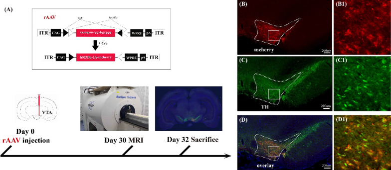
Fig. 1.
Experimental procedure for chemogenetic activation of VTA neurons and immunohistochemical fluorescence imaging. A rAAV encoding the cre-dependent DREADD receptor rAAV-DIO-hM3D(Gq)-mCherry was injected into the right VTA of DAT-cre transgenic rats and an experimental design shows a timeline including the virus injection, behavioral test, and MRI detection. B–D Confirmation of rAAV-infected dopaminergic neurons in the VTA; B the cre-dependent neurons expressing hM3D (Gq) and mCherry proteins (red); C dopaminergic neurons were stained by anti-Tyrosine Hydroxylase (green); D illustration of the virus-infected dopaminergic neurons (yellow) by merging the results of the virus labeling (B) and immunohistochemistry (C). Panels B1, C1, D1 are higher-magnification images of boxed regions in B–D. The nuclei were stained blue by DAPI, and the scale bar: 200 μm

